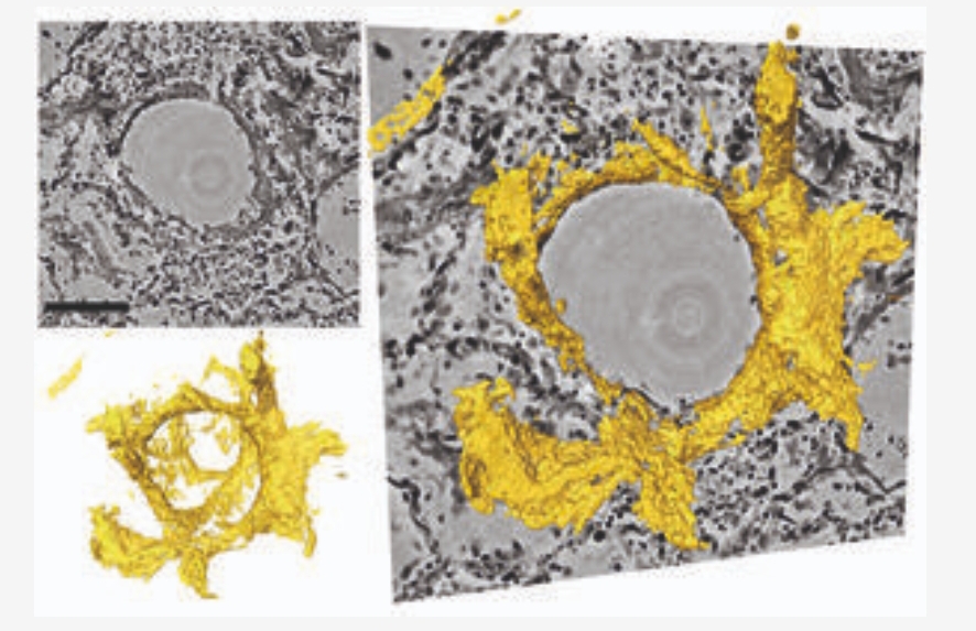
Photo: Tim Salditt, Marina Eckerman
Physicists at the University of Göttingen, together with pathologists and lung specialists at the Medical University of Hannover, have developed a 3D imaging technique that enables h...
Read More

Physicists at the University of Göttingen, together with pathologists and lung specialists at the Medical University of Hannover, have developed a 3D imaging technique that enables h...
Read More
Recent Comments