


. Credit: Michihiro Toritsuka from Fujita Health University School of Medicine, Japan")
Autism spectrum disorder (ASD) is a complex neurodevelopmental condition in which affected individuals experience difficulties in social communication and exhibit restricted, repetitive patterns of behavior or interests.
A growing body of research suggests that neurobiological changes, particularly abnormalities in dendritic spines, tiny protrusions on nerve cells where synapses form, may be a hallmark of ASD...
Read More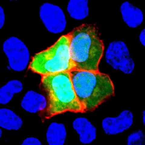


Recent Comments