Epigenetics tagged posts

Epigenetic changes regulate gene expression, but what regulates epigenetics?

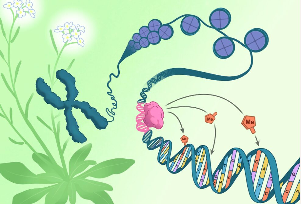
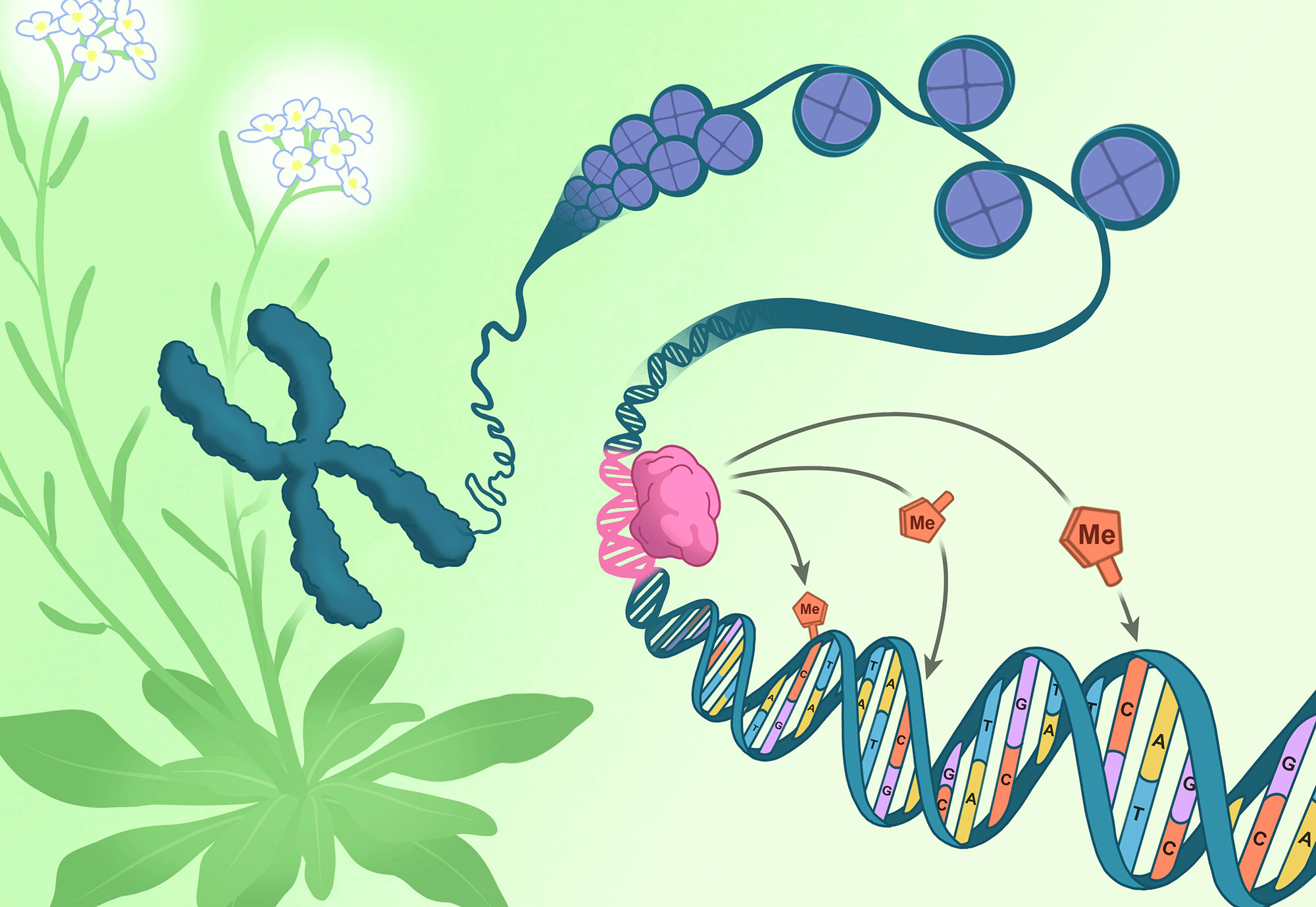
A chromosome pulled from the flowers of Arabidopsis thaliana (green and white) unspools to reveal DNA (blue) coiled around packaging-proteins called histones (purple). The direction of epigenetic changes by genetic features begins as the RIM transcription factor (pink) docks on a corresponding DNA sequence (pink). Once docked, the RIM transcription factor directs methylation machinery to tack methyl groups (orange) onto specific nearby cytosines (orange).
Click here for a high-resolution image.
Credit: Salk Institute

All the cells in an organism have the exact same genetic sequence. What differs across cell types is their epigenetics—meticulously placed chemical tags that influence which genes are expressed in each cell...

Read More

Cancer researchers identify the ‘switch’ that allows intestinal cells to regenerate after injury

Cancer researchers identify the 'switch' that allows intestinal cells to regenerate after injury
Suppressing H3K36 methylation induces the accumulation of abnormal secretory cells. Credit: Nature Cell Biology (2025). DOI: 10.1038/s41556-024-01580-y

Researchers from the University of Colorado Cancer Center have solved a cellular mystery that may lead to better therapies for colorectal and other types of cancer.

Peter Dempsey, Ph.D., professor of pediatrics–developmental biology in the CU School of Medicine, and Justin Brumbaugh, Ph.D., assistant professor of molecular, cellular, and developmental biology at CU Boulder, recently published a paper in the journal Nature Cell Biology showing the importance of the H3K36 methylation process in regulating plasticity and regeneration in intestinal cells.

“The intestine has an enormous ability to regenerate itself after injury, an...

Read More

There’s more to Genes than DNA: How Mum and Dad add something Extra, just for you

Embryos
A normal 4-day-old mouse embryo (L) and an embryo of the same age that has been manipulated to contain maternal chromosomes only (parthenogenote). At this stage, the embryos (blastocysts) appear similar, but the parthenogenote will soon die, underscoring the importance of inheriting imprinted genes from both parents. Different cell types are stained green or red. Credit: Dr Maki Asami.

Biologists at the Universities of Bath and Vienna have discovered 71 new ‘imprinted’ genes in the mouse genome, a finding that takes them a step closer to unravelling some of the mysteries of epigenetics — an area of science that describes how genes are switched on (and off) in different cells, at different stages in development and adulthood.

To understand the importance of imprinted genes to inherit...

Read More

Unexpected Role for Epigenetic Enzymes in Cancer

KDM5s are involved in selection of polyA sites. (A) Western blot and (B) RT-qPCR analyses of MCF7 cells treated with dimethyl sulfoxide (DMSO) or 10 μM KDM5-C70 for 3 days. The ratio of 3′UTR to CDS for CCND1 mRNA was plotted. Error bars represent SEM for biological triplicate experiments. (C) Western blot and (D) RT-qPCR analyses of HeLa cells treated with DMSO or 10 μM KDM5-C70 for 3 days. The ratio of 3′UTR to CDS for DICER1 mRNA was plotted. Error bars represent SEM for biological triplicate experiments. **P < 0.01. (E) RT-qPCR analysis of HeLa/iCas9-c1 cells transduced with lentiviruses carrying single-guide RNAs against KDM5A, KDM5B, KDM5C, or nontargeting control. The ratio of 3′UTR to CDS for DICER1 mRNA was plotted. KO, knockout. Error bars represent SEM for biological triplicate experiments. *P < 0.05; **P < 0.01. (F) Working model for KDM5 involvement in APA. KDM5 recruits the polyA machinery to nascent RNA to modulate polyA site choices. Demethylation or hydroxylation of certain subunits of the polyA machinery by KDM5 also contributes to selection of the polyA sites. The polyA sites (dashes) are noted on the nascent transcript.

KDM5s are involved in selection of polyA sites. (A) Western blot and (B) RT-qPCR analyses of MCF7 cells treated with dimethyl sulfoxide (DMSO) or 10 μM KDM5-C70 for 3 days. The ratio of 3′UTR to CDS for CCND1 mRNA was plotted. Error bars represent SEM for biological triplicate experiments. (C) Western blot and (D) RT-qPCR analyses of HeLa cells treated with DMSO or 10 μM KDM5-C70 for 3 days. The ratio of 3′UTR to CDS for DICER1 mRNA was plotted. Error bars represent SEM for biological triplicate experiments. **P < 0.01. (E) RT-qPCR analysis of HeLa/iCas9-c1 cells transduced with lentiviruses carrying single-guide RNAs against KDM5A, KDM5B, KDM5C, or nontargeting control. The ratio of 3′UTR to CDS for DICER1 mRNA was plotted. KO, knockout...

Read More