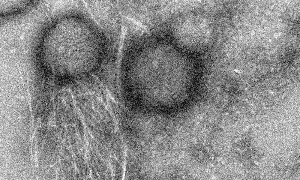
The peptide uperin 3.5 is secreted by the Australian toadlet’s skin. When exposed to bacterial membranes, it rapidly changes its structure and transforms into a deadly antimicrobial weapon. The pictures were taken using a transmission electron microscope (TEM) in the Electron Microscopy Centers in the Technion Department of Materials Science and Engineering and in the Department of Chemical Engineering. The cross-α atomic structure was determined by data collected at the ESRF synchrotron. Credit: Nir Salinas/Technion
Researchers have discovered remarkable molecular properties of an antimicrobial peptide from the skin of the Australian toadlet. The discovery could inspire the development of novel synthetic drugs to combat bacterial infections.
Biologists from The University of Manchester have explained for the first time why having a good night’s sleep really could prepare us for the rigours of the day ahead. The study in mice and published in Nature Cell Biology, shows how the body clock mechanism boosts our ability to maintain our bodies when we are most active.
And because we know the body clock is less precise as we age, the discovery, argues lead author Professor Karl Kadler, may one day help unlock some of the mysteries of aging.
The discovery throws fascinating light on the body’s extracellular matrix -which provides structural and biochemical support to cells in the form of connective tissue such as bone, skin, tendon and cartilage.
Over half our body weight is matrix, and half of this is collagen – and sci...
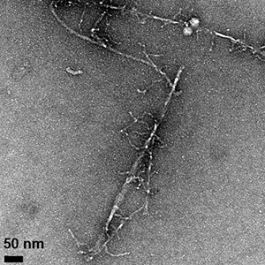
Fibrils formed by the aggregation of the amyloid beta protein can be seen in these transmission electron microscope images, which show differences in fibril morphology between the normal protein (above) and an altered protein with one amino acid replaced by its mirror image. The altered protein also forms fibrils more slowly and is more toxic to cells. (Image credit: Warner et al., CEJ 2016)
Subtle change to amyloid beta protein affects its aggregation behavior, stabilizes an intermediate form with enhanced toxicity. Much of the research on Alzheimer’s disease has focused on the amyloid beta protein, which clumps together into sticky fibrils that form deposits in the brains of people with the disease...


Recent Comments