


As we age, our brains typically undergo a slow process of atrophy, causing less robust communication between various brain regions, which leads to declining memory and other cognitive functions. But a rare group of older individuals called “superagers” have been shown to learn and recall novel information as well as a 25-year-old. Investigators from Massachusetts General Hospital (MGH) have now identified the brain activity that underlies superagers’ superior memory. “This is the first time we have images of the function of superagers’ brains as they actively learn and remember new information,” says Alexandra Touroutoglou, Ph.D...
Read More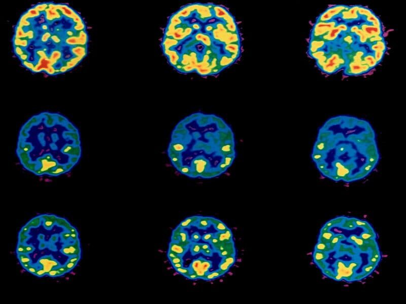


Recent Comments