Glial cells tagged posts

Key Mechanisms identified for Regeneration of Neurons

neuron
Credit: Pixabay/CC0 Public Domain

Neurological disorders, such as trauma, stroke, epilepsy, and various neurodegenerative diseases, often lead to the permanent loss of neurons, causing significant impairments in brain function. Current treatment options are limited, primarily due to the challenge of replacing lost neurons.

Direct neuronal reprogramming, a complex procedure that involves changing the function of one type of cell into another, offers a promising strategy.

In cell culture and in living organisms, glial cells—the non-neuronal cells in the central nervous system—have been successfully transformed into functional neurons. However, the processes involved in this reprogramming are complex and require further understanding...

Read More

Researchers identify a Molecule Critical to Functional Brain Rejuvenation

diagram showing the impact of TET1 in young and old mice
In young adult mice (left), TET1 is active in oligodendroglial cells especially after injury and this leads to new myelin formation and healthy brain function. In old mice (right), the age-related decline of TET1 levels impairs the ability of oligodendroglial cells to form functional new myelin. The authors are currently investigating whether increasing TET1 levels in older mice could rejuvenate the oligodendroglial cells and restore their regenerative functions.

The discovery could have important implications for the health of aging brains and development of therapies for neurodegenerative diseases. Recent studies suggest that new brain cells are being formed every day in response to injury, physical exercise, and mental stimulation...

Read More

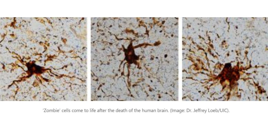
‘Zombie’ Genes? Research shows some Genes Come to Life in the Brain after Death

Post-mortem changes may shed light on important brain studies. In the hours after we die, certain cells in the human brain are still active. Some cells even increase their activity and grow to gargantuan proportions, according to new research from the University of Illinois Chicago.

In a newly published study in the journal Scientific Reports, the UIC researchers analyzed gene expression in fresh brain tissue — which was collected during routine brain surgery — at multiple times after removal to simulate the post-mortem interval and death. They found that gene expression in some cells actually increased after death.

These ‘zombie genes’ — those that increased expression after the post-mortem interval — were specific to one type of cell: inflammatory cells called glial cells...

Read More

Astrocytes eat Connections to maintain Plasticity in Adult Brains

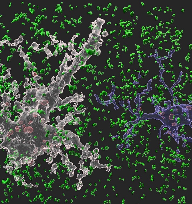
Image: A 3-D animated image showing our synapse phagocytosis reporter in mouse hippocampus. Presynapses in green, astrocytes in white, and microglia in blue. Phagocytosed presynapses by glia were shown in red.
 Image: A 3-D animated image showing our synapse phagocytosis reporter in mouse hippocampus. Presynapses in green, astrocytes in white, and microglia in blue. Phagocytosed presynapses by glia were shown in red.

Developing brains constantly sprout new synapses as they learn and remember. Important connections — the ones that are repeatedly introduced, such as how to avoid danger — are nurtured and reinforced, while connections deemed unnecessary are pruned away. Adult brains undergo similar pruning, but it was unclear how or why synapses in the adult brain get eliminated.

Now, a team of researchers based in Korea has found the mechanism underlying plasticity and, potentially, neurological disorders in adult brains. They published their findings on December 23 in Nature.

“Our findi...

Read More