


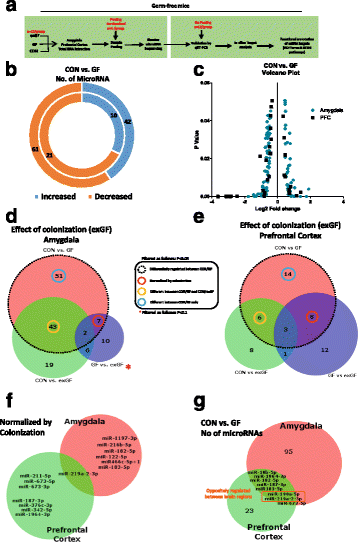
GF male mice display dysregulated network of miRNA expression in the amygdala and PFC. a Schematic representation of experimental design. b Donut plot representing the number of increased and decreased miRNA in the amygdala (outer plot) and PFC (inner plot) when comparing CON vs GF mice. c Volcano plot representing fold change against significance (P < 0.05) between CON and GF mice in the amygdala and PFC. d Venn diagram reporting overlapping differentially regulated miRNA between all three experimental groups in the amygdala representing the effect of colonization of GF mice on miRNAs. e Represents the impact of colonization of GF mice in the PFC. f Number of miRNAs by name that are normalized by colonization and common in both brain regions...


Recent Comments