influenza infection tagged posts

Memory Killer T cells are primed in the Spleen during Influenza infection

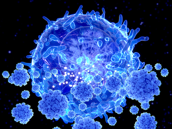
Illustration of a T lymphocyte, or T cell, white blood cells targeting SARS-CoV-2 coronavirus particles. T lymphocytes are components of the body's immune system. Helper T cells stimulate other immune cells to act against a pathogen, whereas killer T cells target and destroy infected cells themselves. SARS-CoV-2 emerged in Wuhan, China, in December 2019, and causes a mild respiratory illness (covid-19) that can develop into pneumonia and be fatal in some cases.
This finding upends the long-held paradigm that priming during lung infections takes place only in the draining lymph nodes, and it will be key to developing more efficient vaccinations and therapies for respiratory challenges.

CD8+ T cells – known as “killer” T cells – are the assassins of the immune system. Once they are primed, they seek out and destroy other cells that are infected with virus or cells that are cancerous.

Priming involves dendritic cells – sentinels of the immune system. In an influenza infection in the lungs, for example, lung-migratory dendritic cells capture a piece of the viral antigen, and then migrate out of the lung to the place where naïve T cells reside, to present that antigen to the CD8+ T cells. This primes the T cells to know which cells to attack.

...Read More

Protein that could Improve Symptoms and Reduce Mortality in Flu

Structural characterization of retrocyclin. (a) CD spectrum demonstrating the similarity in structure between retrocyclin and RTD-1, both at 0.5 mg/ml in a 1:1 mixture of trifluoroethanol in PBS at pH 7.4. (b) A hypothetical model of retrocyclin made by templating its sequence on the backbone of a similar peptide from porcine neutrophils, protegrin-1 (PDB accession code: 1PG1). (c) A cartoon version of b, wherein arginines are black, cysteines are gray, and the other residues are identified by single letter code. (d) A similar cartoon of rhesus RTD-1, indicating the similarity in structure with retrocyclin.

Structural characterization of retrocyclin. (a) CD spectrum demonstrating the similarity in structure between retrocyclin and RTD-1, both at 0.5 mg/ml in a 1:1 mixture of trifluoroethanol in PBS at pH 7.4. (b) A hypothetical model of retrocyclin made by templating its sequence on the backbone of a similar peptide from porcine neutrophils, protegrin-1 (PDB accession code: 1PG1). (c) A cartoon version of b, wherein arginines are black, cysteines are gray, and the other residues are identified by single letter code. (d) A similar cartoon of rhesus RTD-1, indicating the similarity in structure with retrocyclin.

New research in the Journal of Leukocyte Biology identifies potential protective agent against flu infection...

Read More