


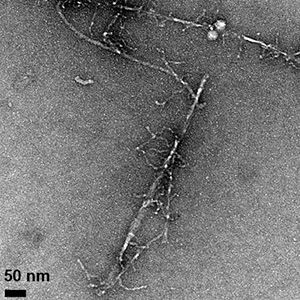
Fibrils formed by the aggregation of the amyloid beta protein can be seen in these transmission electron microscope images, which show differences in fibril morphology between the normal protein (above) and an altered protein with one amino acid replaced by its mirror image. The altered protein also forms fibrils more slowly and is more toxic to cells. (Image credit: Warner et al., CEJ 2016)
Subtle change to amyloid beta protein affects its aggregation behavior, stabilizes an intermediate form with enhanced toxicity. Much of the research on Alzheimer’s disease has focused on the amyloid beta protein, which clumps together into sticky fibrils that form deposits in the brains of people with the disease...
Read More

Recent Comments