

Artificial intelligence (AI) systems are computational models that can learn to identify patterns in data, make accurate predictions or generate content (e.g., texts, images, videos or sound recordings). These models can reliably complete various tasks and are now also used to carry out research rooted in different fields.
Over the past few decades, some AI models have proved promising for the early diagnosis and study of specific diseases or neuropsychiatric conditions. For instance, by analyzing large amounts of brain scans collected using a noninvasive technique known as magnetic resonance imaging (MRI), AI could uncover patterns associated with tumors, strokes and neurodegenerative diseases, which could help to diagnose these conditions.
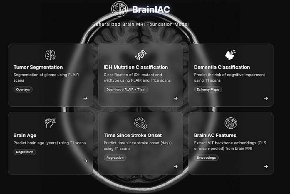
Researchers at Mass General Brigham, Harvard Medical School and other institutes recently developed Brain Imaging Adaptive Core (BrainIAC), a large AI system pre-trained on a vast pool of MRI data that could be adapted to tackle different tasks. This foundation model, presented in a paper published in Nature Neuroscience, was found to outperform many models that were trained to complete specific medical or neuroscience-related tasks.eur
“There is a vast trove of data within the millions of brain MRIs performed each year in the United States,” Benjamin H. Kann, senior author of the paper, told Medical Xpress. “Typically, these scans are analyzed by humans for a particular reason, but this only scratches the surface of the story that these scans might tell us about our patients. With AI and advanced computational imaging techniques, we can unlock much more information from these scans than ever before—which may lead to potent, clinically useful ways to track a variety of acute and chronic conditions, from stroke, to cancer, to dementia, as well as predict future risks for patients.”
The BrainIAC foundation model
BrainIAC, the generic model developed by Kann and his colleagues, was pre-trained on a total of 48,965 brain imaging scans collected using MRI. The model was trained via an approach known as self-supervised learning, which allows models to learn patterns from mostly unlabeled data.
Via this extensive training, the model acquired information about the structure of the human brain and patterns that underpin its organization. Following its initial pre-training, the model can be adapted to detect or study the progression of specific diseases and neuropsychiatric conditions from a patient’s MRI scans.
“This ‘pre-training,’ which uses something called contrastive learning, sets it apart from most MRI AI algorithms that simply train a model to predict a single thing,” explained Kann. “Using this core baseline knowledge, the tool can then be adapted to identify various brain diseases, determine their severity, and predict future risks from these diseases.”
As part of their study, the researchers adapted their model and assessed its ability to detect various conditions, including Alzheimer’s disease, autism, dementia, brain tumors, Parkinson’s disease and strokes. Remarkably, they found that their model could predict most of these conditions with good accuracy and could easily adapt to each of them, requiring little additional training.
“When comparing this model to other models trained on single tasks, the model sometimes needed up to 10 times less training data to perform equivalently well, and sometimes beyond that, than a trained model designed for a single task,” said Kann. “This means that the model can serve as a baseline for predicting many different types of brain disorders.”
A promising tool for medical and neuroscience research
In the future, the BrainIAC model could be improved further and pre-trained on an even larger pool of MRI data. In addition, it could inspire the development of other foundation AI models trained on other types of imaging data, such as computer tomography (CT) scans, high-resolution microscopy images, retinal images, ultrasound recordings and scans of other parts of specific organs.
“We think that this algorithm could help researchers and clinicians use AI for many different types of brain disorders that were infeasible before due to limited amounts of data available,” said Kann.
The BrainIAC algorithm is open-source and is available online on a dedicated website. Other research groups have already started using the model to study various brain-related conditions, detect their emergence and trace their progression.
“We have already received a lot of interest and are collaborating with various groups to investigate BrainIAC for a variety of brain disorders like Alzheimer’s disease and traumatic injury,” added Kann. https://medicalxpress.com/news/2026-02-foundation-ai-mri-multiple-brain.html


Recent Comments