Category Biology/Biotechnology

Microbes Compete for Nutrients, Affect Metabolism, Development in Mice

Nacho Vivas, lab manager at the Rey Lab, checks on a group of germ-free mice inside a sterile environment. Credit: Bryce Richter/UW-Madison

Nacho Vivas, lab manager at the Rey Lab, checks on a group of germ-free mice inside a sterile environment. Credit: Bryce Richter/UW-Madison

If our microbiome overindulges, we might not have access to the nutrients we need. That’s the suggestion from new research that shows mice that harbor high levels of microbes that eat choline are deprived of this essential nutrient. “Gut bacteria get to use a lot of our food before we do,” says Federico Rey, a professor of bacteriology at the University of Wisconsin-Madison. Then we get their leftovers – or their waste. Compared to mice without choline-hungry bacteria, the choline-starved mice had an increased susceptibility to metabolic diseases and gave birth to pups with biochemical alterations in the brain and that exhibited more anxious behaviors...

Read More

NASA’s Webb Telescope will study Our Solar system’s ‘Ocean Worlds’

Possible spectroscopy results from one of Europa’s water plumes.

Possible spectroscopy results from one of Europa’s water plumes. This is an example of the data the Webb telescope could return. Credits: NASA-GSFC/SVS, Hubble Space Telescope, Stefanie Milam, Geronimo Villanueva This spectra was created using NASA’s Planetary Spectrum Generator

NASA’s James Webb Space Telescope will use its capabilities to study the “ocean worlds” of Jupiter’s moon Europa and Saturn’s moon Enceladus, adding to observations previously made by NASA’s Galileo and Cassini orbiters. The Webb telescope’s observations could also help guide future missions to the icy moons.

Europa and Enceladus are on the Webb telescope’s list of targets chosen by guaranteed time observers, scientists who helped develop the telescope and thus get to be among the first to use it to observe the...

Read More

HIIT releases Endorphins in the Brain

High-intensity interval training triggers endorphin release in the brain. Photo: Turku PET Centre

High-intensity interval training triggers endorphin release in the brain. Photo: Turku PET Centre

Finnish researchers at the University of Turku have revealed that exercise-induced endorphin release in the brain depends on the intensity of the exercise. Endorphin release induced by exercise may be an important mechanism which affects exercise motivation and maintenance of regular physical activity. The popular high-intensity interval training (HIIT) leads to endorphin release in the brain, which might alleviate the physical and emotional stress caused by the high-intensity exercise. A less demanding, traditional one-hour aerobic exercise does not cause similar endorphin release.

In the study, HIIT significantly increased the release of endorphins and other opioid peptides in the brain area...

Read More

New Light on Link between Gut Bacteria and Anxiety

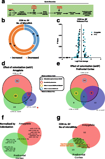
GF male mice display dysregulated network of miRNA expression in the amygdala and PFC. a Schematic representation of experimental design. b Donut plot representing the number of increased and decreased miRNA in the amygdala (outer plot) and PFC (inner plot) when comparing CON vs GF mice. c Volcano plot representing fold change against significance (P < 0.05) between CON and GF mice in the amygdala and PFC. d Venn diagram reporting overlapping differentially regulated miRNA between all three experimental groups in the amygdala representing the effect of colonization of GF mice on miRNAs. e Represents the impact of colonization of GF mice in the PFC. f Number of miRNAs by name that are normalized by colonization and common in both brain regions. g Number of microRNAs that are commonly dysregulated in both brain regions. Highlighted in red are those that are oppositely regulated in both brain regions

GF male mice display dysregulated network of miRNA expression in the amygdala and PFC. a Schematic representation of experimental design. b Donut plot representing the number of increased and decreased miRNA in the amygdala (outer plot) and PFC (inner plot) when comparing CON vs GF mice. c Volcano plot representing fold change against significance (P < 0.05) between CON and GF mice in the amygdala and PFC. d Venn diagram reporting overlapping differentially regulated miRNA between all three experimental groups in the amygdala representing the effect of colonization of GF mice on miRNAs. e Represents the impact of colonization of GF mice in the PFC. f Number of miRNAs by name that are normalized by colonization and common in both brain regions...

Read More