
vision tagged posts

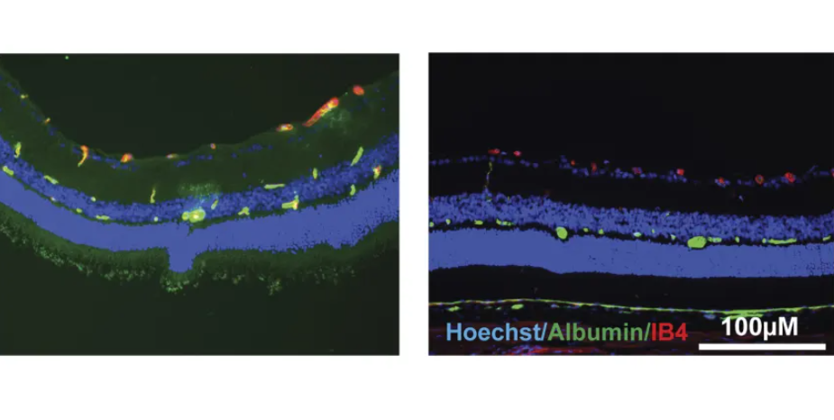
Treatment with norrin helps maintain healthy blood vessels and preserve vision in diabetic rats

Millions of people in the U.S. are affected by diabetic retinopathy, a complication of diabetes that affects the eyes. The condition is caused by damage to the blood vessels in the retina, the light-sensitive tissue at the back of the eye. At first, diabetic retinopathy might not cause any symptoms, or the problems might be mild. However, if the underlying diabetes remains poorly managed, it can lead to blindness.

In a new study, University of Michigan researchers showed that treatment with the molecule norrin can prevent vision loss in diabetic rats. The study is published in the journal Diabetes.

Diabetic retinopathy treatment
Current therapies for diabetic retinopathy target vascular endothelial growth factor, or VEGF, a protein that causes new blood vessels to form.

VEGF a...

Read More
Vision can be rebooted in adults with amblyopia, study suggests

MIT study shows how vision can be rebooted in adults with amblyopia
A new MIT study suggests a strategy for treating adults with amblyopia. Credit: Skitterphoto, CC0, via Wikimedia Commons

Temporarily anesthetizing the retina briefly reverts the activity of the visual system to that observed in early development and enables growth of responses to the amblyopic eye, new research shows.

In the common vision disorder amblyopia, impaired vision in one eye during development causes neural connections in the brain’s visual system to shift toward supporting the other eye, leaving the amblyopic eye less capable even after the original impairment is corrected. Current interventions are only effective during infancy and early childhood while the neural connections are still being formed.

But a new study in mice by neuroscientists in The Picower Institute f...

Read More

Seeing Better by Looking Away

Lead author
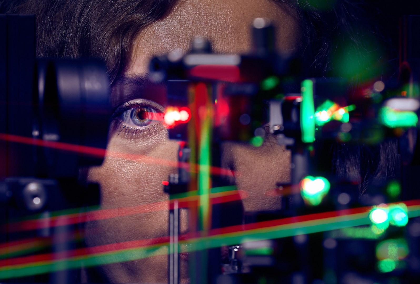
Lead author – Jenny L. Reiniger taking measurements on the laser ophthalmoscope© Volker Lannert/Uni Bonn

A study suggests that we fixate slightly away from the retinal optimum to see better. When we fixate an object, its image does not appear at the place where photoreceptors are packed most densely. Instead, its position is shifted slightly nasally and upwards from the cellular peak. This is shown in a recent study conducted at the University of Bonn (Germany), published in the journal Current Biology. The researchers observed such offsets in both eyes of 20 healthy subjects, and speculate that the underlying fixation behavior improves overall vision.

We like to think of the eye as a camera, but the analogy falls short if we look at the distribution of light sensitive cells — phot...

Read More