

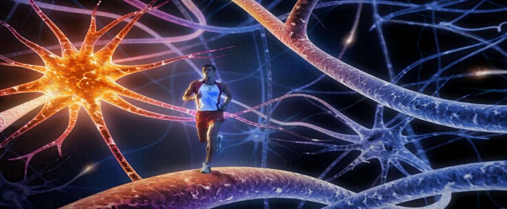


HIIT releases Endorphins in the Brain

High-intensity interval training triggers endorphin release in the brain. Photo: Turku PET Centre

High-intensity interval training triggers endorphin release in the brain. Photo: Turku PET Centre

Finnish researchers at the University of Turku have revealed that exercise-induced endorphin release in the brain depends on the intensity of the exercise. Endorphin release induced by exercise may be an important mechanism which affects exercise motivation and maintenance of regular physical activity. The popular high-intensity interval training (HIIT) leads to endorphin release in the brain, which might alleviate the physical and emotional stress caused by the high-intensity exercise. A less demanding, traditional one-hour aerobic exercise does not cause similar endorphin release.

In the study, HIIT significantly increased the release of endorphins and other opioid peptides in the brain area...

Read More
New Light on Link between Gut Bacteria and Anxiety

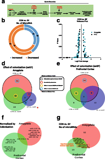
GF male mice display dysregulated network of miRNA expression in the amygdala and PFC. a Schematic representation of experimental design. b Donut plot representing the number of increased and decreased miRNA in the amygdala (outer plot) and PFC (inner plot) when comparing CON vs GF mice. c Volcano plot representing fold change against significance (P < 0.05) between CON and GF mice in the amygdala and PFC. d Venn diagram reporting overlapping differentially regulated miRNA between all three experimental groups in the amygdala representing the effect of colonization of GF mice on miRNAs. e Represents the impact of colonization of GF mice in the PFC. f Number of miRNAs by name that are normalized by colonization and common in both brain regions. g Number of microRNAs that are commonly dysregulated in both brain regions. Highlighted in red are those that are oppositely regulated in both brain regions

GF male mice display dysregulated network of miRNA expression in the amygdala and PFC. a Schematic representation of experimental design. b Donut plot representing the number of increased and decreased miRNA in the amygdala (outer plot) and PFC (inner plot) when comparing CON vs GF mice. c Volcano plot representing fold change against significance (P < 0.05) between CON and GF mice in the amygdala and PFC. d Venn diagram reporting overlapping differentially regulated miRNA between all three experimental groups in the amygdala representing the effect of colonization of GF mice on miRNAs. e Represents the impact of colonization of GF mice in the PFC. f Number of miRNAs by name that are normalized by colonization and common in both brain regions...

Read More

Carbon Nanotube Pores developed to Exclude Salt from Seawater

An artist's depiction of the promise of carbon nanotube porins for desalination. The image depicts a stylized carbon nanotube pipe that delivers clean desalinated water from the ocean to a kitchen tap. Credit: Image by Ryan Chen/LLNL

An artist’s depiction of the promise of carbon nanotube porins for desalination. The image depicts a stylized carbon nanotube pipe that delivers clean desalinated water from the ocean to a kitchen tap. Credit: Image by Ryan Chen/LLNL

Lawrence Livermore scientists, in collaboration with Northeastern University have developed a saltwater purification device. The team also found that water permeability in carbon nanotubes (CNTs) with diameters smaller than a nanometer (0.8 nm) exceeds that of wider CNTs by an order of magnitude. The nanotubes, hollow structures made of carbon atoms in a unique arrangement, are more than 50,000 times thinner than a human hair...

Read More

No Batteries Required: Energy-harvesting Yarns generate Electricity

Coiled carbon nanotube yarns, created at the University of Texas at Dallas and imaged here with a scanning electron microscope, generate electrical energy when stretched or twisted. Credit: University of Texas at Dallas

Coiled carbon nanotube yarns, created at the University of Texas at Dallas and imaged here with a scanning electron microscope, generate electrical energy when stretched or twisted. Credit: University of Texas at Dallas

An international team led by scientists at The University of Texas at Dallas and Hanyang University in South Korea has developed high-tech yarns that generate electricity when they are stretched or twisted. In a study published in the Aug. 25 issue of the journal Science, researchers describe “twistron” yarns and their possible applications, such as harvesting energy from the motion of ocean waves or from temperature fluctuations. When sewn into a shirt, these yarns served as a self-powered breathing monitor.

“The easiest way to think of twistron harvesters is, you have a p...

Read More