

A recent USC study provides new information about why SARS-CoV-2, the virus behind the COVID-19 pandemic, may elicit mild symptoms at first but then, for a subset of patients, turn potentially fatal a week or so after infection. The researchers showed that distinct stages of illness correspond with the coronavirus acting differently in two different populations of cells.
The study, published in Nature Cell Biology, may provide a roadmap for addressing cytokine storms and other excessive immune reactions that drive serious COVID-19.
The team found that when SARS-CoV-2 infects its first-phase targets, cells in the lining of the lung, two viral proteins circulate within those cells—one that works to activate the immune system and a second that, paradoxically, blocks that signal, resulting in little or no inflammation.
The team also discovered a second pathway the virus sometimes takes to enter immune cells. This alternative pathway both stunts the virus’s ability to reproduce and prevents the production of the second immune signal-braking protein. The first protein is then able to spur rampant inflammation linked to severe symptoms.
“There are two stages that work through different signaling pathways,” said Rongfu Wang, Ph.D., a professor of pediatrics and medicine at the Keck School of Medicine of USC. “With the normal pathway, everything goes normally, and the virus replicates. When the immune cells pick up the virus, replication is defective, but it produces a lot of inflammatory signaling molecules called cytokines.”
In lab experiments, the Keck School of Medicine scientists identified a drug that quelled inflammation in human immune cells infected with SARS-CoV-2 and reduced symptoms in mice, suggesting that it may be possible to prevent deadly cytokine storms.
Exploring inflammation in COVID-19
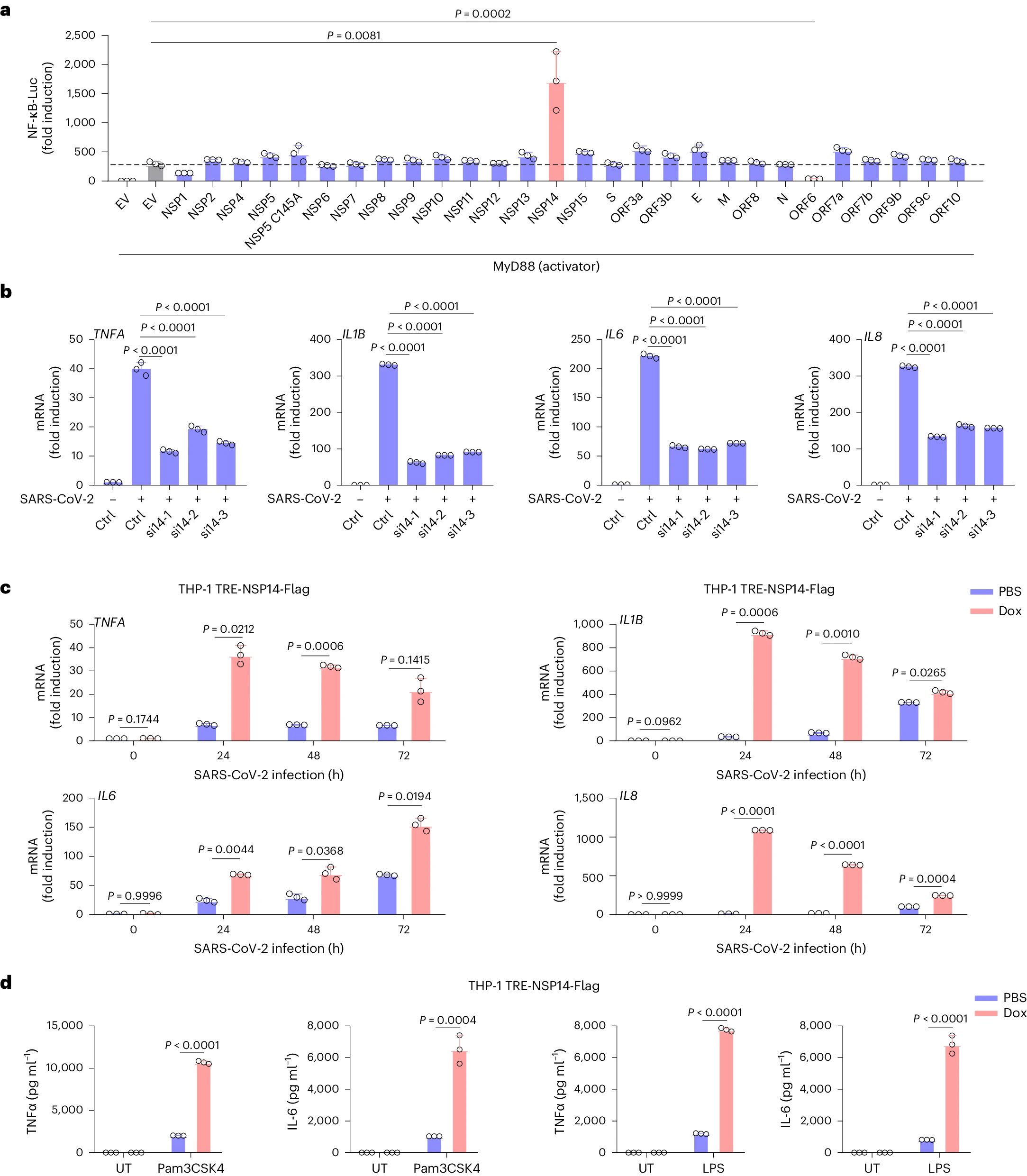
Wang and his colleagues began with two related core questions: Why is so little inflammation seen with SARS-CoV-2 in lung cells? And, for the people who don’t recover in the first seven to 10 days, why do the symptoms get so much worse? The researchers started by screening all SARS-CoV-2 proteins to see which ones regulate the production of cytokines. Their search yielded two results: a protein called NSP14, which causes inflammation, and another called ORF6, which quells it.
The team followed up on this finding with an exhaustive series of experiments encompassing protein-on-protein reactions, human and animal cell lines, lab models, and tissue and fluid samples from COVID-19 patients.
This is the picture that emerged: When the coronavirus enters a cell of the lung lining through its known entry point, the ACE2 protein on the cell’s surface, both NSP14 and ORF6, are produced within the cell. ORF6 acts as a guardian at the membrane surrounding the cell nucleus, effectively silencing the inflammatory signal.
“In this population of lung cells, ORF6 overrides NSP14 so that no cytokines are produced,” said Wang, who is also the Endowed Chair in Cell Therapy Research at Children’s Hospital Los Angeles. “Even if NSP14 takes action, ORF6 doesn’t let it get into the cell nucleus. The door is closed, so it’s completely blocked for cytokine production.”
Discoveries about the coronavirus’s direct effect on the body’s natural defenses
While it has been suspected that SARS-CoV-2 affects immune cells, to date, there hasn’t been a clear view of how this works. The Keck School research team shed substantial light on this topic.
They mapped out the process for how SARS-CoV-2 interacts with immune cells, which generally lack ACE2 proteins on their surface. Wang and his colleagues found that immune cells take the virus inside through a membrane protein called TLR1, which acts as a sort of virus sensor as part of the body’s first line of defense. Instead of the coronavirus’s well-known spike protein latching onto these cells, SARS-CoV-2 connects using one of the envelope proteins on its surface.
The nature of this separate entrance prevents the production of ORF6, eliminating the anti-inflammatory signal. The pro-inflammatory protein NSP14 then drives immune cells to send out cytokines that can cause the serious, sometimes fatal symptoms seen in later-stage COVID-19.
In one of the most striking pieces of evidence, human immune cells that were genetically engineered so that they lacked the TLR1 protein showed no signs of inflammatory response when infected with SARS-CoV-2. When two other types of cells that normally lack ACE2 were altered so that they had TLR1 on their surface and then exposed to SARS-CoV-2, the coronavirus was able to gain entry.
The study not only expanded our understanding of COVID-19 infection but also offered new clues about how to manage the later, serious stage of the disease. In experiments with mice, treating COVID-19 with a drug that blocks TLR1 led to longer survival and less weight loss compared to an untreated control group.
The findings’ larger implications suggest that early-stage COVID-19 is best addressed by attempting to stymie viral replication, while later-stage therapy should focus on taming inflammation.
“Inflammation is a major issue in many diseases,” Wang said. “If you can control inflammation, you can control a lot of diseases. Depending on the situation, you either want to activate the immune system when something’s wrong or calm it when there’s nothing else wrong.” https://keck.usc.edu/news/covid-19-research-new-details-about-potentially-deadly-inflammation-revealed-in-usc-study/:~:text=Press%20Release-,COVID%2D19%20research%3A%20New%20details%20about%20potentially%20deadly%20inflammation%20revealed,is%20associated%20with%20severe%20inflammation


Recent Comments